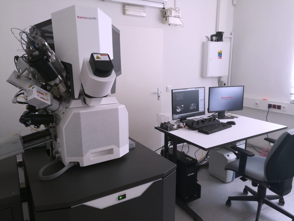
A new microscope will allow RCPTM to rank with top European scientific centres
A new scanning electron microscopy system [SEM] with a focused ion beam [FIB], which recently enriched the RCPTM infrastructure, will allow a faster and more effective sample analysis. From an instrumental perspective, the combination of methods used to date with the new will allow RCPTM to rank among top scientific centres worldwide.
Besides imaging nanoscale structures, the equipment enables nanoscale work with samples, providing information not only about surfaces of materials [2D imaging] but also particular depth [3D] profiles. SEM/FIB can be used to create so-called lamellas for TEM imaging and analysis of samples that are too thick for primary analysis by transmission electron microscopy [TEM, HRTEM]. Moreover, it does not change important properties of samples. “The goal of preparing these lamellas is to look inside the studied materials and image their inner structure. The combination of SEM/FIB will allow accurate localization of the target site or area from which the desired lamella should be made,” explained Štěpán Kment from the Photoectrochemistry group.

Development of new materials constitutes a major part of RCPTM’s research activities, often in the form of complex hybrid nanostructures. The desired functional properties of these advanced systems are usually related to localized phenomena or interactions, e.g. interface structure between layers, boundaries of heterogeneous crystalline grains, form of binding, and interaction of nanoparticles. The structural and to some extent chemical nature of the localized phenomena can be studied by TEM and primarily HRTEM methods.
“The new microscope will allow high-level investigation of the inner structure of materials that could not be studied previously, despite the centre having state-of-the-art TEM and HRTEM microscopes. From an instrumental perspective, the combination of the mentioned technologies [SEM/FIB, TEM, and HRTEM] will allow RCPTM to join the top ranks of European scientific centres in capacity to characterize structurally and chemically complex nanomaterials, thereby increasing its global competitiveness.” said Kment.
RCPTM Director Radek Zbořil mentioned other benefits: “A complex system of electron microscopes in combination with UHV STM techniques [ultra-high vacuum scanning tunnelling microscope] gives us a unique chance of imaging surfaces of nanomaterials at the atomic level in detail, along with structures, properties and reactions of molecules.”
This purchase worth 19.6 million CZK was made with the grant project Nanotechnologies for Future [CZ.02.1.01/0.0/0.0/16_019/0000754] solved within the call Excellence in Research; Operational Programme Research, Development and Education


