 Riley, KE; Hobza P: “On the Importance and Origin of Aromatic Interactions in Chemistry and Biodisciplines”, ACC. CHEM. RES. (2013) 46, 927-936.
DOI: 10.1021/ar300083h, IF=24.348.
Abstract – Aromatic systems contain both σ- and π-electrons, which in turn constitute σ- and π-molecular orbitals (MOs). In discussing the properties of these systems, researchers typically refer to the highest occupied and lowest unoccupied MOs, which are π MOs. The characteristic properties of aromatic systems, such as their low ionization potentials and electron affinities, high polarizabilities and stabilities, and small band gaps (in spectroscopy called the N → V1 space), can easily be explained based on their electronic structure. These one-electron properties point to characteristic features of how aromatic systems interact with each other.
Unlike hydrogen bonding systems, which primarily interact through electrostatic forces, complexes containing aromatic systems, especially aromatic stacked pairs, are predominantly stabilized by dispersion attraction. The stabilization energy in the benzene dimer is rather small (2.5 kcal/mol) but strengthens with heteroatom substitution. The stacked interaction of aromatic nucleic acid bases is greater than 10 kcal/mol, and for the most stable stacked pair, guanine and cytosine, it reaches approximately 17 kcal/mol. Although these values do not equal the planar H-bonded interactions of these bases (29 kcal/mol), stacking in DNA is more frequent than H-bonding and, unlike H-bonding, is not significantly weakened when passing from the gas phase to a water environment.
Consequently, the stacking of aromatic systems represents the leading stabilization energy contribution in biomacromolecules and in related nanosystems. Therefore stacking (dispersion) interactions predominantly determine the double helical structure of DNA, which underlies its storage and transfer of genetic information. Similarly, dispersion is the dominant contributor to attractive interactions involving aromatic amino acids within the hydrophobic core of a protein, which is critical for folding.
Therefore, understanding the nature of aromatic interactions, which depend greatly on quantum mechanical (QM) calculations, is of key importance in biomolecular science. This Account shows that accurate binding energies for aromatic complexes should be based on computations made at the (estimated) CCSD(T)/complete basis set limit (CBS) level of theory. This method is the least computationally intensive one that can give accurate stabilization energies for all common classes of noncovalent interactions (aromatic–aromatic, H-bonding, ionic, halogen bonding, charge-transfer, etc.). These results allow for direct comparison of binding energies between different interaction types. Conclusions based on lower-level QM calculations should be considered with care.
 Lazar, P; Zhang, S; Safarova, K; Li, Q; Froning, JP; Granatier, J; Hobza, P; Zboril, R; Besenbacher, F; Dong, MD; Otyepka, M: “Quantification of the Interaction Forces between Metals and Graphene by Quantum Chemical Calculations and Dynamic Force Measurements under Ambient Conditions”, ACS NANO (2013) 7, 1646-1651.
DOI: 10.1021/nn305608a, IF=12.033.
Abstract – The two-dimensional material graphene has numerous potential applications in nano(opto)electronics, which inevitably involve metal graphene interfaces.Theoretical approaches have been employed to examine metal graphene interfaces, but experimental evidence is currently lacking. Here, we combine atomic force microscopy (AFM) based dynamic force measurements and density functional theory calculations to quantify the interaction between metal-coated AFM tips and graphene under ambient conditions. The results show that copper has the strongest affinity to graphene among the studied metals (Cu, Ag, Au, Pt, Si), which has important implications for the construction of a new generation of electronic devices. Observed differences in the nature of the metal–graphene bonding are well reproduced by the calculations, which included nonlocal Hartree–Fock exchange and van der Waals effects.
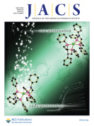 Lazar, P; Karlicky, F; Jurecka, P; Kocman, M; Otyepkova, E; Safarova, K; Otyepka, M; : “Adsorption of Small Organic Molecules on Graphene”, J. AM. CHEM. SOC., (2013) 135 (16), 6372-6377.
DOI: 10.1021/ja403162r, IF=11.444.
Abstract – We present a combined experimental and theoretical quantification of the adsorption enthalpies of seven organic molecules (acetone, acetonitrile, dichloromethane, ethanol, ethyl acetate, hexane, and toluene) on graphene. Adsorption enthalpies were measured by inverse gas chromatography and ranged from −5.9 kcal/mol for dichloromethane to −13.5 kcal/mol for toluene. The strength of interaction between graphene and the organic molecules was estimated by density functional theory (PBE, B97D, M06-2X, and optB88-vdW), wave function theory (MP2, SCS(MI)-MP2, MP2.5, MP2.X, and CCSD(T)), and empirical calculations (OPLS-AA) using two graphene models: coronene and infinite graphene. Symmetry-adapted perturbation theory calculations indicated that the interactions were governed by London dispersive forces (amounting to 60% of attractive interactions), even for the polar molecules. The results also showed that the adsorption enthalpies were largely controlled by the interaction energy. Adsorption enthalpies obtained from ab initio molecular dynamics employing non-local optB88-vdW functional were in excellent agreement with the experimental data, indicating that the functional can cover physical phenomena behind adsorption of organic molecules on graphene sufficiently well.
 Bartkiewicz, K; Lemr, K; Černoch, A; Soubusta, J; Miranowicz, A: “Experimental Eavesdropping Based on Optimal Quantum Cloning“, PHYS. REV. Lett. (2013) 110, 173601.DOI: 10.1103/PhysRevLett.110.173601, IF=7.728.
Abstract – The security of quantum cryptography is guaranteed by the no-cloning theorem, which implies that an eavesdropper copying transmitted qubits in unknown states causes their disturbance. Nevertheless, in real cryptographic systems some level of disturbance has to be allowed to cover, e.g., transmission losses. An eavesdropper can attack such systems by replacing a noisy channel by a better one and by performing approximate cloning of transmitted qubits which disturb them but below the noise level assumed by legitimate users. We experimentally demonstrate such symmetric individual eavesdropping on the quantum key distribution protocols of Bennett and Brassard (BB84) and the trine-state spherical code of Renes (R04) with two-level probes prepared using a recently developed photonic multifunctional quantum cloner [Lemr et al., Phys. Rev. A 85 050307(R) (2012)]. We demonstrated that our optimal cloning device with high-success rate makes the eavesdropping possible by hiding it in usual transmission losses. We believe that this experiment can stimulate the quest for other operational applications of quantum cloning.
 RCPTM members (Hamal, P.; Nozka, L.) have contributed to a number of ATLAS Collaboration articles that form a significant part among the most valued publications of the Centre. As of April 2013, 2 Phys. Rev. Lett. (IF=7.728) papers and 5 J. High Energy Phys. (IF=6.220) have been published in 2013 from the areas of the search for the Z-boson and dark matter candidates.
 Krajewski, S.; Prucek, R.; Panacek, A.; Avci-Adali, M.; Nolte, A.; Straub, A.; Zboril, R., Wendel, H. P.; Kvitek, L.: “Hemocompatibility evaluation of different silver nanoparticle concentrations employing a modified Chandler-loop in vitro assai on human blood“, ACTA BIOMATER. (2013) 9, 7460–7468.
DOI: 10.1016/j.bios.2013.01.043, IF=5.684.
Abstract – Due to their antibacterial effects, the use of silver nanoparticles (AgNPs) in a great variety of medical applications like coatings of medical devices has increased markedly in the last few years. However, blood in contact with AgNPs may induce adverse effects, thereby altering hemostatic functions. The objective of this study was to investigate the hemocompatibility of AgNPs in whole blood. Human whole blood (n = 6) was treated with different AgNPs concentrations (1, 3 and 30 mg l−1) or with saline/blank solutions as controls before being circulated in an in vitro Chandler-loop model for 60 min at 37 °C. Before and after circulation, various hematologic markers were investigated. Based on the hematologic parameters measured, no profound changes were observed in the groups treated with AgNP concentrations of 1 or 3 mg l−1. AgNP concentrations of 30 mg l−1 induced hemolysis of erythrocytes and α-granule secretion in platelets, increased CD11b expression on granulocytes, increased coagulation markers thrombin–antithrombin-III complex, kallikrein-like and FXIIa-like activities as well as complementing cascade activation. Overall, we provide for the first time a comprehensive evaluation including all hematologic parameters required to reliably assess the hemocompatibility of AgNPs. We strongly recommend integrating these hemocompatibility tests to preclinical test procedures prior to in vivo application of new AgNP-based therapies.
 Adegboyega, NF; Sharma, VK; Siskova, K; Zboril, R; Sohn, M; Schultz, BJ; Banerjee, S: “Interactions of Aqueous Ag+ with Fulvic Acids: Mechanisms of Silver Nanoparticle Formation and Investigation of Stability”, ENVIRON. SCI. TECHNOL. (2013) 47, 757-764,
DOI: 10.1021/es302305f, IF=5.481.
Abstract – This study investigated the possible natural formation of silver nanoparticles (AgNPs) in Ag
+–fulvic acid (FA) solutions under various environmentally relevant conditions (temperature, pH, and UV light). Increase in temperature (24–90 °C) and pH (6.1–9.0) of Ag
+–Suwannee River fulvic acid (SRFA) solutions accelerated the appearance of the characteristic surface plasmon resonance (SPR) of AgNPs. The rate of AgNP formation via reduction of Ag
+ in the presence of different FAs (SRFA, Pahokee Peat fulvic acid, PPFA, Nordic lake fulvic acid, NLFA) and Suwannee River humic acid (SRHA) followed the order NLFA > SRHA > PPFA > SRFA. This order was found to be related to the free radical content of the acids, which was consistent with the proposed mechanism. The same order of AgNP growth was seen upon UV light illumination of Ag
+–FA and Ag
+–HA mixtures in moderately hard reconstituted water (MHRW). Stability studies of AgNPs, formed from the interactions of Ag
+–SRFA, over a period of several months showed that these AgNPs were highly stable with SPR peak reductions of only 15%. Transmission electron microscopy (TEM) and dynamic light scattering (DLS) measurements revealed bimodal particle size distributions of aged AgNPs. The stable AgNPs formed through the reduction of Ag
+ by fulvic and humic acid fractions of natural organic matter in the environment may be transported over significant distances and might also influence the overall bioavailability and ecotoxicity of AgNPs.
 Markova, Z; Siskova, K; Filip, J; Cuda, J; Kolar, M; Safarova, K; Medrik, I; Zboril, R: “Air Stable Magnetic Bimetallic Fe-Ag Nanoparticles for Advanced Antimicrobial Treatment and Phosphorus”, ENVIRON. SCI. TECHNOL. (2013).
DOI: 10.1021/es304693g, IF=5.481.
Abstract – We report on new magnetic bimetallic Fe–Ag nanoparticles (NPs) which exhibit significant antibacterial and antifungal activities against a variety of microorganisms including disease causing pathogens, as well as prolonged action and high efficiency of phosphorus removal. The preparation of these multifunctional hybrids, based on direct reduction of silver ions by commercially available zerovalent iron nanoparticles (nZVI) is fast, simple, feasible in a large scale with a controllable silver NP content and size. The microscopic observations (transmission electron microscopy, scanning electron microscopy/electron diffraction spectroscopy) and phase analyses (X-ray diffraction, Mössbauer spectroscopy) reveal the formation of Fe
3O
4/γ-FeOOH double shell on a “redox” active nZVI surface. This shell is probably responsible for high stability of magnetic bimetallic Fe–Ag NPs during storage in air. Silver NPs, ranging between 10 and 30 nm depending on the initial concentration of AgNO
3, are firmly bound to Fe NPs, which prevents their release even during a long-term sonication. Taking into account the possibility of easy magnetic separation of the novel bimetallic Fe–Ag NPs, they represent a highly promising material for advanced antimicrobial and reductive water treatment technologies.
 Prucek, R; Tuček, J; Kolařík, J; Filip, J; Marušák, Z; Sharma, VK; Zbořil, R:“Ferrate(VI)-Induced Arsenite and Arsenate Removal by In-Situ Structural Incorporation into Magnetic Iron(III) Oxide Nanoparticles”, ENVIRON. SCI. TECHNOL. (2013) 47, 3283–3292.
DOI: 10.1021/es3042719, IF=5.481.
Abstract – We report the first example of arsenite and arsenate removal from water by incorporation of arsenic into the structure of nanocrystalline iron(III) oxide. Specifically, we show the capability to trap arsenic into the crystal structure of γ-Fe
2O
3 nanoparticles that are in situ formed during treatment of arsenic-bearing water with ferrate(VI). In water, decomposition of potassium ferrate(VI) yields nanoparticles having core–shell nanoarchitecture with a γ-Fe
2O
3 core and a γ-FeOOH shell. High-resolution X-ray photoelectron spectroscopy and in-field
57Fe Mössbauer spectroscopy give unambiguous evidence that a significant portion of arsenic is embedded in the tetrahedral sites of the γ-Fe
2O
3 spinel structure. Microscopic observations also demonstrate the principal effect of As doping on crystal growth as reflected by considerably reduced average particle size and narrower size distribution of the “in-situ” sample with the embedded arsenic compared to the “ex-situ” sample with arsenic exclusively sorbed on the iron oxide nanoparticle surface. Generally, presented results highlight ferrate(VI) as one of the most promising candidates for advanced technologies of arsenic treatment mainly due to its environmentally friendly character, in situ applicability for treatment of both arsenites and arsenates, and contrary to all known competitive technologies, firmly bound part of arsenic preventing its leaching back to the environment. Moreover, As-containing γ-Fe
2O
3 nanoparticles are strongly magnetic allowing their separation from the environment by application of an external magnet.
 Baratella, D; Magro, M; Sinigaglia, G; Zboril, R; Salviulo, G; Vianello, F: “A glucose biosensor based on surface active maghemite nanoparticles“, BIOSENS. BIOELECTRON. (2013) 45, 13–18. DOI: 10.1016/j.actbio.2013.03.016, IF=6.451.
Abstract – A simple carbon paste (CP) electrode, modified with novel maghemite (γ-Fe2O3) nanoparticles, called SAMNs (suface active maghemite nanoparticles) and characterized by a mean diameter of about 10 nm, has been developed. The electrode catalyzes the electro-reduction of hydrogen peroxide at low applied potentials (−0.1 V vs SCE). In order to improve the electrocatalytic properties of the modified electrode an ionic liquid, namely 1-butyl-3-methylimidazolium hexafluorophosphate (BMIM-PF6), was introduced. At −0.1 V, the sensitivity of the SAMN–BMIM-PF6–CP electrode was 206.51 nA μM−1 cm−2, with a detection limit (S/N=3) of 0.8 μM, in the 0–1.5 mM H2O2 concentration range. Furthermore, glucose oxidase was immobilized on the surface of maghemite nanoparticles as a monomolecular layer, by a bridge constituted of rhodamine B isothiocyanate, leading to a fluorescent, magnetic drivable nanocatalyst, containing 10±2 enzyme molecules per nanoparticle. The resulting enzyme electrode presents a linear calibration curve toward glucose in solution in the concentration range of 0–1.5 mM glucose, characterized by a sensitivity of 45.85 nA μM−1 cm−2 and a detection limit (S/N=3) of 0.9 μM. The storage stability of the system was evaluated and a half-life of 2 months was calculated, if the electrode is stored at 4 °C in buffer. The present work demonstrates the feasibility of these surface active maghemite nanoparticles as efficient hydrogen peroxide electro-catalyst, which can be easily coupled to hydrogen peroxide producing enzymes in order to develop oxidase based reagentless biosensor devices.
 Riley, KE; Hobza P: “On the Importance and Origin of Aromatic Interactions in Chemistry and Biodisciplines”, ACC. CHEM. RES. (2013) 46, 927-936.
DOI: 10.1021/ar300083h, IF=24.348.
 Lazar, P; Zhang, S; Safarova, K; Li, Q; Froning, JP; Granatier, J; Hobza, P; Zboril, R; Besenbacher, F; Dong, MD; Otyepka, M: “Quantification of the Interaction Forces between Metals and Graphene by Quantum Chemical Calculations and Dynamic Force Measurements under Ambient Conditions”, ACS NANO (2013) 7, 1646-1651.
DOI: 10.1021/nn305608a, IF=12.033.
 Lazar, P; Karlicky, F; Jurecka, P; Kocman, M; Otyepkova, E; Safarova, K; Otyepka, M; : “Adsorption of Small Organic Molecules on Graphene”, J. AM. CHEM. SOC., (2013) 135 (16), 6372-6377.
DOI: 10.1021/ja403162r, IF=11.444.
 Bartkiewicz, K; Lemr, K; Černoch, A; Soubusta, J; Miranowicz, A: “Experimental Eavesdropping Based on Optimal Quantum Cloning“, PHYS. REV. Lett. (2013) 110, 173601.DOI: 10.1103/PhysRevLett.110.173601, IF=7.728.
 RCPTM members (Hamal, P.; Nozka, L.) have contributed to a number of ATLAS Collaboration articles that form a significant part among the most valued publications of the Centre. As of April 2013, 2 Phys. Rev. Lett. (IF=7.728) papers and 5 J. High Energy Phys. (IF=6.220) have been published in 2013 from the areas of the search for the Z-boson and dark matter candidates.
 Krajewski, S.; Prucek, R.; Panacek, A.; Avci-Adali, M.; Nolte, A.; Straub, A.; Zboril, R., Wendel, H. P.; Kvitek, L.: “Hemocompatibility evaluation of different silver nanoparticle concentrations employing a modified Chandler-loop in vitro assai on human blood“, ACTA BIOMATER. (2013) 9, 7460–7468.
DOI: 10.1016/j.bios.2013.01.043, IF=5.684.
 Adegboyega, NF; Sharma, VK; Siskova, K; Zboril, R; Sohn, M; Schultz, BJ; Banerjee, S: “Interactions of Aqueous Ag+ with Fulvic Acids: Mechanisms of Silver Nanoparticle Formation and Investigation of Stability”, ENVIRON. SCI. TECHNOL. (2013) 47, 757-764,
DOI: 10.1021/es302305f, IF=5.481.
 Markova, Z; Siskova, K; Filip, J; Cuda, J; Kolar, M; Safarova, K; Medrik, I; Zboril, R: “Air Stable Magnetic Bimetallic Fe-Ag Nanoparticles for Advanced Antimicrobial Treatment and Phosphorus”, ENVIRON. SCI. TECHNOL. (2013).
DOI: 10.1021/es304693g, IF=5.481.
 Prucek, R; Tuček, J; Kolařík, J; Filip, J; Marušák, Z; Sharma, VK; Zbořil, R:“Ferrate(VI)-Induced Arsenite and Arsenate Removal by In-Situ Structural Incorporation into Magnetic Iron(III) Oxide Nanoparticles”, ENVIRON. SCI. TECHNOL. (2013) 47, 3283–3292.
DOI: 10.1021/es3042719, IF=5.481.
 Baratella, D; Magro, M; Sinigaglia, G; Zboril, R; Salviulo, G; Vianello, F: “A glucose biosensor based on surface active maghemite nanoparticles“, BIOSENS. BIOELECTRON. (2013) 45, 13–18. DOI: 10.1016/j.actbio.2013.03.016, IF=6.451.