
Carbon dots assist in human stem cell imaging
Carbon quantum dots, which are highly photostable and fluorescent, were discovered 15 years ago. Since then, they have been widely used as fluorescent labels for in vitro bioimaging, i.e. fluorescently marking various cells, substances and/or structures in cells. They are also being increasingly used as intravenously administered fluorescent contrast agents for in vivo imaging of healthy and diseased tissues. This application exploits their biocompatibility, derived from their simple element composition (predominately carbon, oxygen, and nitrogen). RCPTM scientists have recently attempted to combine the labelling and contrast uses for fluorescently monitoring stem cells in a living organism after their subcutaneous transplantation and intravenous application. They proved to be efficient contrast agents for in vitro imaging of human stem cells, and for the first time intravenously applied mesenchymal stem cells labelled with carbon dots were detected in a mouse tumour.
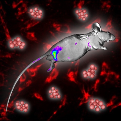
Carbon quantum dots were also used as fluorescent labels for semi-quantitative detection of the transplanted cells and confirmation of the stem cells’ migration to the afflicted inflammatory/tumour site, so-called stem cell homing. Thus, carbon dots may play important roles in the major uses of stem cells: targeted therapy and regenerative medicine. This work follows several recent studies by scientists from Olomouc on uses of carbon dots in biomedicine (e.g. Bao X. et al. Light-Sci. Appl. 7, 91, 2018; Holá K. et al. ACS Nano 11, 12402–12410, 2017; Kalytchuk S. et al. ACS Nano 11, 1432-1442, 2017).